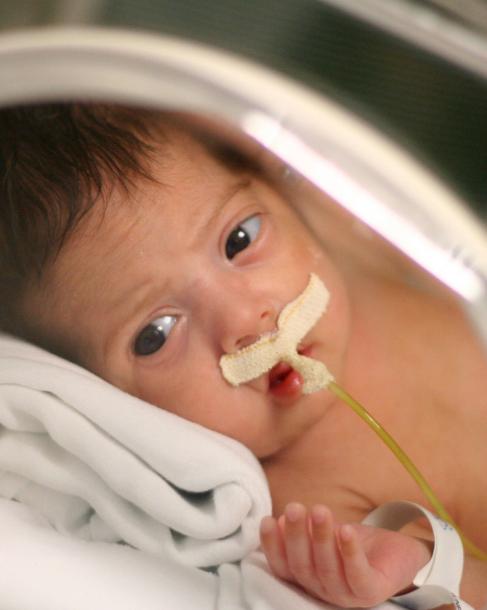
Craniosynostosis is a birth defect in which the bones of a baby’s skull fuse too early, leading to abnormal head shape and, in some cases, unhealthy pressure on the baby’s growing brain.
In normal development, sections at the top of the skull remain separated for the first two decades of life, with flexible joints called sutures between them. There are also two larger gaps called fontanels at the top of the skull, one just behind the forehead and the other at the back of the skull. These separations between sections of the skull allow the brain to grow and the head to gradually change shape in infancy.
In craniosynostosis, babies are born with one or more of these sutures or fontanels closed. What should be separated sections of the skull join prematurely, constraining growth. When this constraint changes the shape of the skull significantly enough to impair normal development, surgical correction can separate the skull’s prematurely joined sections and reshape the skull for healthy growth.
The doctors at UCI Plastic Surgery are leaders in the field of cosmetic and reconstructive surgery, with special expertise in treating children. Through their teaching and lecturing roles at UCI, they stay current with state-of-the-art techniques and technology. With their advanced technical skills, they consistently produce better, more natural-looking results for patients of all ages.
To learn more about how surgery can correct craniosynostosis and other craniofacial development problems, contact UCI Plastic Surgery to schedule a consultation at one of our three locations: in Orange, Costa Mesa, and Tustin, CA.
Types of Craniosynostosis
Craniosynostosis can occur on its own, with no other birth anomalies (non-syndromic craniosynostosis), or as part of a syndrome or cluster of anomalies. Syndromes associated with craniosynostosis include Apert syndrome, Crouzon syndrome, Muenke syndrome, Pfeiffer syndrome, and Saethre-Chotzen syndrome.
Doctors define different forms of craniosynostosis by which of the skull’s sutures close too early in a baby’s development. Each has a different effect on the shape of the head as the baby grows.
Sagittal synostosis
Occurs when the suture running along the top of the head closes too early. This condition is the most common form of craniosynostosis, resulting in a long and narrow head shape.
Unicoronal synostosis
The second most common form of this disorder – occurs when one of the sutures on the side of the head closes too early. This phenomenon can cause the forehead to appear flattened on the affected side of the head. The eye socket may also be raised and the nose bent toward that side of the head.
Bicoronal synostosis
Occurs when the sutures on both sides of the head close too early. The result is an abnormally broad and short head shape with a tall forehead.
Lambdoid synostosis
Occurs when either or both of the sutures in the back of the head close too early. The result is a flattening of the back of the head and displacement of one or both ears.
Metopic synostosis
Occurs when the suture in the front of the head, which runs from the nose up through the forehead, closes too early. The result is a narrowing of the front of the head and a widening of the rear of the head.
Who Can Diagnose Craniosynostosis?
Signs of craniosynostosis are usually present at birth as an unusually shaped head. The head shape typically becomes more abnormal as the baby grows.
Not every baby with a misshapen head has craniosynostosis. Lying in one position can cause a baby’s head to change shape — to flatten in the back or on one side, for example. It’s often possible to correct these problems by changing the baby’s sleeping position or, in some cases, with helmet therapy.
Specialists such as pediatric neurosurgeons or plastic and reconstructive surgeons can accurately diagnose craniosynostosis. A physical exam is the first step, often followed by imaging studies, such as a CT scan, to show where skull sutures have closed. If a doctor suspects a syndrome is responsible for the condition, they may recommend genetic testing.
Craniosynostosis Treatments
Mild cases of craniosynostosis may not need treatment.
If the condition is causing a noticeable and permanent deformity to the head or face, surgery can give the child a more normal appearance and avert later problems with social life and self-esteem.
If the condition is constraining the growth of the child’s brain, surgery will be necessary to prevent developmental delays or cognitive impairment. Surgery may also prevent vision problems if the skull malformation is affecting the eyes.
The timing and type of surgery will depend on the type and severity of the child’s craniosynostosis and whether it is occurring in combination with other abnormalities as part of a genetic syndrome.
Start Your Journey Today!
UCI Plastic Surgery is a leader in the field of cosmetic surgery. Each of our specialists is highly knowledgeable, trained, and committed to bringing our patients the latest advancements in the field. Learn how our experts can help you obtain industry-leading results by scheduling a consultation today.
Schedule A ConsultationHow Do Surgeons Correct Craniosynostosis?
Various surgical techniques can correct different types of malformation at several stages of a child’s development. The surgical team usually includes a specialist in plastic and reconstructive surgery of the head and face, plus a specialist in brain surgery.
The goal of the surgery is to open the closed suture (the joint between two sections of the skull that should remain flexible in infants) to enable the skull to grow into a more normal shape without putting pressure on the developing brain. There are two main approaches to this.
- Endoscopic surgery can be ideal for babies less than three to six months of age. A surgeon makes small incisions through which to insert a camera and surgical instruments to remove the closed suture in the skull. This separation of two joined sections of the skull is called strip craniectomy.
- Open surgery is for babies over six months old, who have multiple suture involvement, or when more extensive reshaping of the skull is required. The surgeon makes an incision, removes the closed suture, reshapes the skull as needed, and places absorbable plates and screws to hold the newly shaped bones in position.
Beyond those incision approaches, the surgeon will choose from a wide range of techniques to correct the child’s specific skull malformation.
- The procedure may involve placing stainless steel springs to correct sagittal synostosis, expand the space between the separated sections of the skull, and give the brain more space for growth.
- With other forms of craniosynostosis, a surgeon may place cranial distractors to gradually expand areas of the skull. These external devices affix to points on newly separated skull sections, and require periodic adjustment to expand the skull and enable the development of new bone.
- Fronto-orbital advancement can correct misshapen bones around the eye and forehead. This procedure involves removing and recontouring the forehead bone and upper eye bones.
- Cranial vault remodeling and reconstruction reshapes the skull when it is deformed or when more than one suture has closed prematurely. A surgeon removes and reshapes the malformed sections of skull, and then secures it back into place with absorbable plates and/or sutures.
- As a child grows older, they may need more than one surgery to expand and reshape parts of the skull. Once the skull has fully matured, facial contouring surgery can refine the balance of the facial features.
What to Expect in Recovery
Children who have surgery to correct craniosynostosis will recover for one to four days in the hospital. When the staff at UCI Plastic Surgery clears your child to go home, they will provide specific instructions for recovery, including guidance on how to care for dressings, manage your child’s pain, and bathe your child. Your child will be able to resume a regular diet and normal activity immediately on release from the hospital. To minimize the risk of injury, we suggest affixing cushions on the edges and corners of low furniture such as coffee tables.
Your doctor will schedule follow-up appointments to track your child’s progress in healing and will be available to respond to your questions.
Schedule a Consultation in Orange County
Do you have a child with craniosynostosis in Southern California? Contact us today to schedule a consultation and find out how surgical repair can set your child on track for normal development. We’ll answer your questions and explain the surgical approach we recommend, based on your child’s unique needs. We have three Orange County offices: in Orange, Costa Mesa, and Tustin, CA.